
Epilepsy is a chronic disabling disorder affecting over 1% of the population who suffer higher rates of physical, mental and social disability and associated costs of over $10,000 per year. Neurosurgery can be curative in many cases. Surgery can have cognitive risks, however: up to 41% patients still suffer language decline after temporal lobe surgery1 and 44% suffer verbal memory loss. There is a dearth of validated, reproducible methods to simply and accurately predict patient outcome. My research mission is to develop standardized, evidence-based protocols to accurately predict patients’ real-world functioning after epilepsy surgery.
fMRI for mapping the brain’s language areas
Clinical interpretation of language fMRI. To obtain an accurate picture of how clinical fMRI is used and interpreted, we surveyed 82 epilepsy surgical program directors and clinicians. In addition to using fMRI to identify the language dominant hemisphere, 44% reported relying on fMRI to guide surgical margins to preserve language function. Further, at most 21% sought to map other known language regions beyond Broca’s and Wernicke’s areas. Instances of unpredicted language decline were reported by 17%, and 54% reported cases of unexpected preservation of function. None of these cases had been published.
Technical execution of fMRI. Separately, there are no clear technical guidelines covering the tasks, training, and analytic approaches required for presurgical fMRI, and the standards of practice in each of these areas was unknown. We surveyed 63 analysts across epilepsy surgical programs and found heterogeneity in all aspects of fMRI. Multiple variants of over 15 protocols were in use, and neither of the two best-validated protocols were used by more than 10% of programs. Many sites reported completing fMRI without professionals trained in neuropsychology (64%) and radiology (34%)–disciplines with complementary skills fundamental to this method.
These data show that the interpretation and execution of fMRI in the clinic varies from the best available evidence, a fact that will lead to varying accuracy in predicting post-surgical decline. Separately, they suggest that in spite of a lack of evidence, many clinicians already use fMRI maps to guide surgical margins and that the majority only recognize two language areas when they do so. This is significant as at least four further language regions are both identifiable using fMRI and are associated with language deficits when removed or directly stimulated.
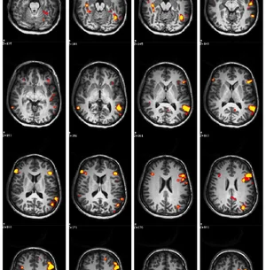
Mapping of language cortex beyond Broca’s and Wernicke’s areas. The above data suggests a clinical need for fMRI to identify critical language areas. We defined a new approach to estimate the location of six known language areas, including Broca’s & Wernicke’s areas, Exner’s Area, Supplementary Speech Area, Angular Gyrus, and the Basal Temporal Language Area. This method relies on a set of three different language tasks and a trained clinician selecting and combining data from the tasks to identify these regions. In 22 epilepsy patients, this method was reliable when used by different clinicians (78% overlap) and identified the Wada-defined language-dominant hemisphere with accuracy equivalent to the best published methods (85% of cases).12 Further, activation consistent with all six language regions consistently and more often than an automated analysis of the same data.
A reliable, validated, freely-available multilingual battery (ongoing). The above work will have limited impact on patients’ lives if it cannot be reproduced, or is available only in English. Further, the above data showed that over half of surgical programs rely on closed (and often unvalidated) commercial software packages (52%) rather than freely-available published alternatives, and that 68% of US epilepsy programs use unvalidated in-house translations of tasks (most often in Spanish). We have provided the above tasks11 free for download on this site, and in over fifteen different languages (further translations pending). These protocols have been downloaded forty times to date, and have led to developing collaborations with Portuguese, Greek and South African epilepsy programs.
Vision - optic radiations mapping
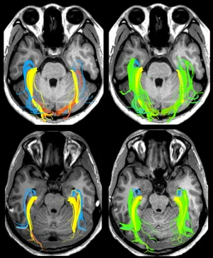
Another goal of surgical planning is the mapping of the visual system’s optic radiations. This vital structure is extremely hard to map with MRI due to its circuitous route. Researchers have largely focused on improving MRI acquisition methods to better map the structure. This study showed that a prominent idea in the field, that the original work of Meyer in 1908 showed that the radiations include three structures, is incorrect. It also showed that existing methods for finding this structure are highly variable, and showed that a combination of these works more effectively. This work was supported by, and completed under the guidance of, Professor Simon Warfield, PhD within his Computational Radiology Laboratory.
Spatial memory and the temporal lobes
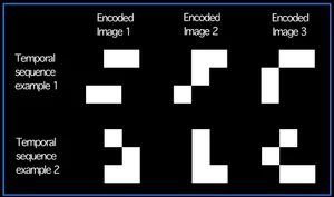
A further goal in planning surgical treatment in epilepsy is understanding which brain structures in the temporal lobe support memory, and how well they are functioning. This allows the effects of different forms of surgery to be predicted during planning. The key to this has been seen as understanding which hemisphere’s mesial temporal lobe (MTL) is critical in verbal memory. Curiously, we remain unable to map memory for this purpose using fMRI. In this study we studied which temporal lobe structures are important in encoding the “space” and “time” of experience; for instance, so that you can remember the order of events that occurred over a few minutes, and where everything (people, objects, etc.) where during this time. The results from this functional MRI study provided further evidence that a key function of the temporal lobe is gluing information together for storage in memory, and suggest that different areas in the middle portion of the temporal lobe (“medial temporal lobe”) achieve this in different ways.
Publications
Publications demonstrating the above, and others, are viewable here: